Skin cancer is one of the most preventable—and most treatable—cancers when caught early, yet many cases are discovered later than they need to be. Proactive skin checks and professional mole screening turn uncertainty into clarity: you learn what’s normal for your skin, spot changes sooner, and take practical steps to lower risk without living in fear.
The Importance of Early Detection: Understanding Skin Cancer and Its Types
Early detection matters in skin cancer for a simple biological reason: most skin cancers grow outward and then deeper over time. The deeper certain cancers extend into the skin, the more access they gain to blood vessels and lymphatic channels—pathways that can allow spread. Catching suspicious lesions while they’re small and superficial often means simpler treatment, smaller scars, and far better outcomes.
Skin cancer is not one disease. Understanding the main types helps you know what you’re looking for during self-checks and why professionals take specific lesions seriously.
Basal cell carcinoma (BCC) is the most common type. It arises from basal cells in the epidermis and is strongly linked to cumulative ultraviolet (UV) exposure. BCC usually grows slowly and rarely spreads to distant sites, but it can become locally destructive if ignored—eroding into cartilage or bone, particularly on the nose, ears, and around the eyes.
Typical appearances include a pearly or shiny bump, a persistent sore that doesn’t heal, a scaly patch, or a scar-like area that seems to appear without an injury. Some BCCs are subtle and look like a faint pink patch or a slightly depressed area. If you’ve ever wondered, “Why won’t this spot just go away?”—that’s the type of question that should trigger a check.
Squamous cell carcinoma (SCC) is also common and related to cumulative UV exposure, but it’s more likely than BCC to spread if left untreated—especially SCC on the lips, ears, or in people with weakened immune systems. SCC often presents as a rough, scaly, thickened patch; a firm red bump; or a lesion that crusts, bleeds, or feels tender. Think “persistent and irritated,” particularly on sun-exposed areas like the scalp, face, forearms, and hands.
Melanoma is less common than BCC or SCC but more dangerous because it can spread more rapidly. It begins in melanocytes (pigment-producing cells). The good news is that when melanoma is detected early—before it grows deep—treatment is often straightforward and highly effective. The challenge is that melanoma can be highly variable. It can be dark brown or black, but it can also be tan, red, pink, or even nearly colorless (amelanotic melanoma).
Other skin cancers exist, including Merkel cell carcinoma (rare but aggressive), dermatofibrosarcoma protuberans (usually slow-growing), and cutaneous lymphomas. Most people will never encounter these, but they underscore an important point: not every dangerous lesion looks like a “classic mole.” Any persistent, changing, or unusual spot deserves attention.
Precancerous changes also matter. Actinic keratoses (AKs) are rough, sandpaper-like spots caused by sun damage and can develop into SCC. Treating AKs is a proactive strategy—one that reduces future risk rather than waiting for cancer to appear.
A final reality check: skin cancer risk isn’t limited to people who burn easily or spend all day outdoors. Intermittent intense sun exposure, tanning bed use, childhood sunburns, fair skin, a large number of moles, family history, and immune suppression all play roles. And melanoma can occur in people with darker skin tones too—often in less sun-exposed areas like palms, soles, and under nails—making targeted awareness essential.
Recognizing the Signs: How to Perform Effective Self-Examinations and Identify Warning Signs
A professional skin exam is important, but it can’t replace what happens in between visits. Self-exams help you recognize your personal baseline and notice changes early. The goal isn’t to diagnose yourself—it’s to catch “new, changing, or unusual” so you can bring it to a clinician promptly.
How often should you check? Monthly is a practical standard for many adults, especially if you have risk factors like a history of sunburns, numerous moles, or prior skin cancer. If you’re low risk, you might do it every 2–3 months—but consistency matters more than perfection.
What you need: good lighting, a full-length mirror, a hand mirror, and something to document what you see (your phone camera or a simple body map). If you have a partner or family member willing to help, areas like the scalp and back become easier.
Step-by-step self-exam (a methodical “head-to-toe” scan):
1) Face and scalp. Check the nose, lips, ears (front and back), and around the eyes. For the scalp, use a comb or hair dryer to part hair in sections. If you have thinning hair or a shaved head, the scalp is a high-risk zone.
2) Hands and arms. Look at the palms, backs of hands, between fingers, and around nails. Then examine wrists, forearms, elbows, and upper arms.
3) Torso. Check the neck, chest, and abdomen. Women should lift the breasts to check underneath. Look at the sides of your body with arms raised.
4) Back and buttocks. Use mirrors for the upper back, lower back, and buttocks. Don’t forget the back of the neck and behind the ears.
5) Legs and feet. Examine the front and back of thighs, shins, calves, ankles, tops of feet, soles, between toes, and the nail beds. Melanoma can appear under a nail as a dark streak that widens, changes, or involves the surrounding skin (a red flag).
What exactly are you looking for? Focus on two categories: “moles” and “non-mole lesions.” Skin cancers don’t always look like moles.
The ABCDE rule for moles (especially for melanoma):
A – Asymmetry: one half doesn’t match the other.
B – Border: irregular, scalloped, or poorly defined edges.
C – Color: multiple colors or uneven distribution (brown, black, red, white, blue).
D – Diameter: often larger than 6 mm (about a pencil eraser), though melanomas can be smaller.
E – Evolving: any change in size, shape, color, elevation—or new symptoms like itching, tenderness, or bleeding.
The “ugly duckling” sign: This is often more useful than ABCDE in real life. Most people’s benign moles look similar to one another—the same “family.” The mole that doesn’t fit the pattern (darker, redder, larger, more irregular, or just different) is the one to watch.
Warning signs beyond moles:
- A sore that doesn’t heal within 3–4 weeks.
- Bleeding with minimal trauma (for example, a spot that bleeds when you towel off).
- A scaly or crusted patch that persists or returns.
- A pearly bump or a pink growth with visible small blood vessels.
- A firm, rough, thickened area that feels like sandpaper.
- Rapid change in any lesion over weeks to months.
Documenting changes without overreacting: Take a clear photo next to a ruler or coin for scale, under good light, from the same distance. Do this again in 4–6 weeks if you’re uncertain—but don’t “monitor” a clearly changing or symptomatic lesion indefinitely. A lesion that’s evolving, bleeding, or markedly different should be evaluated sooner rather than later.
Common beginner confusion: People often worry about perfectly benign features like freckles, cherry angiomas (small red dots), seborrheic keratoses (waxy “stuck-on” growths), or dermatofibromas (firm small bumps often from old insect bites). These can look alarming, but they also tend to be stable over time. The professional’s job is to differentiate what’s harmless from what needs a biopsy—your job is to notice what’s new or changing.
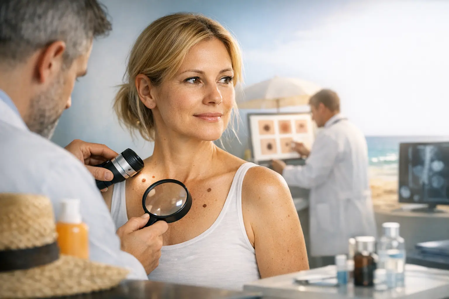
The Role of Professional Screening: What to Expect During a Mole Check and How to Prepare
Self-exams are powerful, but professional screening is where pattern recognition and diagnostic tools come in. Clinicians don’t only look at one mole in isolation—they assess your entire skin, your “mole signature,” and your risk profile. This is particularly valuable if you have many moles, atypical moles, a personal or family history of melanoma, or significant sun exposure history.
What happens during a professional skin exam?
1) Risk assessment and history. Expect questions about sunburns (especially in childhood), tanning bed use, outdoor work or hobbies, previous biopsies, personal history of skin cancer, family history, and immune status. These details change how aggressively clinicians monitor and manage lesions.
2) Full-body skin examination. A thorough exam typically includes scalp, behind the ears, back, buttocks, and feet—areas patients frequently miss. You’ll usually be offered a gown. If you’re uncomfortable, you can discuss what areas will be examined and why.
3) Dermoscopy. Many dermatology practices use a handheld device called a dermatoscope. It uses polarized light and magnification to reveal structures beneath the surface—pigment networks, vessels, and patterns that help distinguish benign from suspicious lesions. Dermoscopy improves accuracy and can reduce unnecessary biopsies while increasing detection of early melanoma.
4) Photography and monitoring (when appropriate). For patients with many moles, clinicians may recommend total body photography and digital dermoscopic monitoring. This isn’t about watching everything forever—it’s about comparing lesions over time. Subtle changes can be detected earlier, and stable lesions can be left alone confidently.
5) Biopsy decisions. If a lesion looks suspicious, clinicians may recommend biopsy. This is not a reason to panic; it’s a diagnostic step. Many biopsies come back benign. But when cancer is found, biopsy is often what enables early, curative treatment.
How to prepare for your appointment:
- Remove nail polish (especially if you can) so nail beds are visible.
- Avoid heavy makeup and consider wearing your hair in a way that allows scalp access.
- Write down concerns in advance: spots that itch, bleed, change, or look different.
- Bring photos if you’ve been tracking a lesion’s evolution.
- Don’t self-treat suspicious spots with acids, freezing kits, or “natural” remedies beforehand; this can delay diagnosis and alter the lesion’s appearance.
How often should you get screened professionally? It depends on risk. People with previous melanoma or multiple atypical moles may be seen every 3–12 months. Average-risk adults may do annual or periodic checks, especially after age 40 or with cumulative sun exposure. If you’re unsure, ask your clinician to define your risk category and recommend an interval based on it.
When to book sooner: a rapidly changing lesion, spontaneous bleeding, a new dark spot after age 30 that looks different from your other moles, a non-healing sore, or any lesion that concerns you enough that you keep thinking about it. Your intuition matters—bring the concern in rather than waiting for the next routine appointment.
Proactive Measures: Lifestyle Adjustments and Preventative Strategies for Skin Health
Screening finds problems; prevention reduces how many problems develop in the first place. UV radiation damages DNA in skin cells. Over time, that damage accumulates, increasing the chance of mutations that lead to cancer. The strategy, then, is to reduce avoidable UV exposure while keeping your approach realistic enough to maintain long-term.
Use sunscreen as a tool, not a loophole. Sunscreen is not permission to roast longer in the sun. It’s one layer of protection in a broader plan.
- Choose broad-spectrum SPF 30+ (higher for prolonged outdoor exposure). Broad-spectrum means UVA and UVB protection.
- Apply enough: most adults need roughly a shot-glass amount to cover the body, and a generous layer for face/neck/ears.
- Reapply every 2 hours and after swimming or heavy sweating, even if labeled “water-resistant.”
- Don’t forget high-miss areas: ears, hairline, back of neck, tops of feet, and hands.
Seek shade strategically. UV intensity peaks when the sun is highest (often 10 a.m. to 4 p.m., depending on location and season). Planning outdoor exercise earlier or later can significantly reduce dose without changing your lifestyle dramatically.
Wear protective clothing. This is one of the most effective and underused strategies—because it doesn’t wear off.
- Wide-brim hat (not just a baseball cap) for ears and neck coverage.
- UV-protective sunglasses to reduce eye damage and protect surrounding skin.
- UPF-rated clothing for long outdoor days; dark, tightly woven fabrics also help.
Avoid tanning beds completely. Indoor tanning devices deliver concentrated UV radiation that accelerates DNA damage and skin aging. If you want a cosmetic tan, choose sunless tanners (dihydroxyacetone-based products). They don’t protect against UV, but they also don’t carry the same carcinogenic UV exposure.
Be mindful around reflective surfaces. Water, sand, snow, and even concrete reflect UV. That’s why skiers and beachgoers can burn quickly—even on cloudy days. Clouds reduce visible brightness more than they reduce UV intensity.
Consider medication and medical factors. Some medications increase sun sensitivity (certain antibiotics, diuretics, retinoids). Immune suppression—whether from organ transplant, autoimmune therapy, or certain conditions—raises skin cancer risk substantially and may warrant more frequent professional screening.
Nourish skin health without overpromising. No supplement replaces sun protection. A diet with adequate antioxidants (from fruits and vegetables), hydration, and avoidance of smoking supports overall skin resilience and healing. But the measurable risk reduction comes primarily from UV behavior changes and early detection.
Make prevention automatic: a realistic routine.
- Keep sunscreen next to your toothbrush so it becomes a morning default.
- Store a travel-sized tube in your bag, car, or gym kit.
- Set a phone reminder to reapply when outdoors for extended periods.
- Schedule a monthly “mole check” on the same date (e.g., first weekend of each month).
Prevention is also psychological: when you know you’re taking practical steps, you’re less likely to avoid checking out of fear. Confidence comes from systems, not guesswork.
Navigating Treatment Options: Understanding Diagnosis, Treatment Pathways, and Follow-Up Care for Skin Cancer
When a clinician is concerned about a lesion, the pathway usually starts with a biopsy. Understanding the process reduces anxiety and helps you participate in decisions.
Biopsy basics:
- Shave biopsy removes the top layers; often used for raised lesions.
- Punch biopsy uses a circular tool to remove a small core; common for inflammatory rashes or certain pigmented lesions.
- Excisional biopsy removes the entire lesion with a margin; often preferred when melanoma is strongly suspected.
Biopsies are typically quick, done under local anesthesia, and followed by simple wound care instructions. The pathology report then guides next steps. Ask for a clear explanation of what was found and what it means for risk and follow-up.
If the diagnosis is basal cell carcinoma: Treatment options depend on location, size, and subtype.
- Surgical excision removes the cancer with a margin of normal skin; widely used and effective.
- Mohs micrographic surgery is especially useful on the face and other cosmetically or functionally sensitive areas. It removes tissue in thin layers and checks margins immediately, preserving healthy skin while achieving high cure rates.
- Curettage and electrodesiccation (scrape and cauterize) may be used for certain low-risk lesions.
- Topical therapies or radiation can be considered in select cases, often when surgery isn’t ideal.
If the diagnosis is squamous cell carcinoma: Many SCCs are treated similarly to BCC, but SCC has a higher potential to spread, so clinicians may be more cautious with margins and follow-up.
- Excision or Mohs surgery are common.
- Evaluation of lymph nodes may be considered for high-risk SCCs (large size, deeper invasion, certain locations, or immune-suppressed patients).
- Actinic keratoses treatment often becomes part of the plan to reduce future SCC risk (e.g., cryotherapy or field treatments for sun-damaged skin).
If the diagnosis is melanoma: Management is more protocol-driven because depth and other histologic features correlate with risk.
- Wide local excision is standard to remove additional tissue around the melanoma site.
- Sentinel lymph node biopsy may be recommended depending on depth and features to assess microscopic spread.
- Additional treatments (immunotherapy, targeted therapy, radiation) may be used for higher-stage disease.
Many people are surprised to learn that early melanoma is often treated with surgery alone. This circles back to why proactive checks matter: early-stage disease usually means simpler treatment and a far better prognosis.
Follow-up care: what “all clear” really means. After any skin cancer diagnosis, your risk of developing another skin cancer increases. That doesn’t mean recurrence is inevitable—it means your skin has demonstrated vulnerability to UV damage or other risk factors. Follow-up appointments are a prevention strategy in themselves.
- Expect regular skin exams at intervals based on your diagnosis and risk.
- Continue monthly self-checks and document new or changing lesions.
- Watch the treatment site for changes, persistent tenderness, new growth, or non-healing areas, and report them.
- Ask for a personalized plan: What should trigger an urgent visit? How often should you be seen? Do you need total body photography?
Scar care and recovery: Proper wound care reduces infection risk and improves cosmetic outcome. Follow your clinician’s instructions closely. If stitches are used, keep follow-up for removal. Once the wound has closed, some clinicians recommend silicone gel or sheets to optimize scar appearance—particularly in high-tension areas.
Common decision point: “Monitor or biopsy?” Monitoring can be appropriate for clearly benign-appearing lesions in low-risk patients, especially with baseline photography. But if a lesion is evolving, symptomatic, or an “ugly duckling,” biopsy is often the responsible choice. When in doubt, ask the clinician to explain the reasoning in plain terms—and what changes would shift the plan.
Conclusion
Proactive skin cancer checks aren’t about turning every freckle into a crisis—they’re about building a clear, repeatable system that catches meaningful change early. Learn the common patterns of BCC, SCC, and melanoma, commit to monthly self-exams that cover the easy-to-miss areas, and treat professional mole screening as a high-value health appointment rather than a cosmetic add-on. Then back it all up with prevention that actually works: consistent sun protection, protective clothing, and zero tanning beds. If something is new, changing, bleeding, or simply doesn’t fit your skin’s usual “mole pattern,” get it evaluated promptly. Early action is the simplest path to peace of mind—and, when needed, the easiest path to effective treatment.

